


CLINICAL GUIDELINES
Background: Osteonecrosis of the femoral head is a progressive disease that can lead to impaired hip function and disability in young patients. Early diagnosis and treatment can help slow the progression of this disease. One of the modern approaches to the treatment of osteonecrosis is core decompression with the introduction of orthobiological products such as bone marrow aspirate concentrate (BMAC).
Aim: Comparison of the results of magnetic resonance imaging of patients diagnosed with osteonecrosis of the femoral head after using two treatment methods: isolated tunneling and tunneling using bone marrow aspirate concentrate.
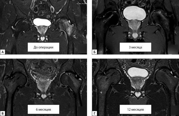
Materials and methods: A prospective cohort non-randomized comparative study was conducted to evaluate the effectiveness of core decompression with the addition of bone marrow aspirate concentrate for the treatment of osteonecrosis of the femoral head. The study involved 108 patients with stages II and IIIA according to the ARCO classification, who were divided into two groups: the main group, which received core decompression withbone marrow aspirate concentrate, and the comparison group, which received only core decompression. The outcome was assessed using MRI (1.5 Tl) to analyze the morphological structure of the hip joints before and after surgery, after 3, 6, and 12 months of follow-up.
Results: In the total sample, at the time of the last control visit, data on 100 (92.6%) patients were available, which made it possible to analyze 168 (91.8%) hip joints. 12 months after surgery, patients with the second stage of the disease in the main group had only two cases of progression (10.3%), while in the comparison group this figure was 18 cases (48.6%), which is significantly higher (p<0.001).
Conclusion: Core decompression with the introduction of bone marrow aspirate concentrate is an effective method of treating osteonecrosis of the femoral head at the ARCO II stage (before collapse) compared with isolated core decompression in the medium term. This method significantly reduces the frequency of disease progression and the need for hip replacement.
Background: Justification. Age-related changes are accompanied by structural disorders not only in the bones of the skeleton, but also in the weakening of sensorimotor systems that regulate the biomechanical functions of the entire musculoskeletal system and in particular the spine. Although visually, the observed balancing disorders are particularly noticeable in the postural functions of balance and the axial skeleton, their quantitative instrumental assessment and diagnosis have not yet been worked out. To date, 3D video gait analysis is a recognized objective research method that allows orthostatics and walking to record and quantify the basic balancing values of the axial skeleton.
Aim: To study the balancing behavior of the axial skeleton in locomotor activity and establish normative statistical guidelines for its balancing characteristics in a three-coordinate space.
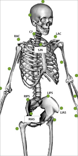
Materials and methods: The study involved men (n=10) and women (n=9) aged 18–40, with an average age of 31.1±1.4 years. Qualisys 7+ optical cameras (8 cameras) with passive marker video capture technology recorded kinematic gait parameters. A specially created program determined the values of dynamic axial balance by the amplitude of the projection of the calculated points from the center of the C7 and S2 vertebrae to the reference plane in the sagittal (GA-SVA) and frontal (GA-CVA) planes and the angle range of the projection of the shoulder and pelvis lines in the horizontal (GA-APA) plane.
Results: The stochastic dynamics of the axial balance of the body analyzed in the study approached the normal distribution function with the lowest level of variation in the horizontal plane. The ranges of coefficients of variation of the axial balance of the body during normal walking "on tiptoes" were: in the horizontal plane (APA) — 7.5%–10.0%; in the sagittal plane SVA — 12.4%–18.6%; in the frontal plane CVA — 12.6%–16.9%.
Conclusion: The statistics of reference values of the dynamic axial balance of the body in the age group of 18–40 years with normal walking and with the functional test "walking on toes" are presented. In the locomotor stochastic hierarchy of biomechanical functions of the axial skeleton, the normalized values of the three-coordinate parameter of axial balance (PAB), should be considered the leading diagnostic guideline in assessing motor pathology, since the distribution of its values is constant and may correspond to the rule of sigma deviations.
REVIEWS
High competitive and training loads in sports are a risk factor for stress fractures in athletes. Vitamin D, which regulates phosphorus-calcium metabolism, affects the condition of the musculoskeletal system. Given the high incidence of vitamin D deficiency in athletes, its correction may have a positive effect on the incidence of injuries in sports. The aim of the review is to assess the effect of vitamin D on bone metabolism in sports based on the results of an analysis of modern scientific publications. The review is based on publications from the PubMed, Google Scholar, Cyberleninka, and eLIBRARY databases. Preference was given to articles in peer-reviewed sources published over the past 10 years. The review emphasizes the relevance of the problem of vitamin D deficiency in sports, highlighting risk factors for the development of its insufficient supply among athletes and its impact on the functioning of the musculoskeletal system. In athletes, the risk of osteopenia is additionally associated with the presence of unfavorable variants of allelic combinations of the genes of the vitamin D receptor and type I collagen, the development of the syndrome of relative energy deficiency in sports. Athletes are recommended to maintain target levels of 25-hydroxyvitamin D (25(OH)D), which have a positive effect on bone remodeling, but specialized recommendations on this issue have not yet been developed. Evaluation of the concentration of 25(OH)D in the blood serum can be recommended for all representatives of the sports subpopulation, especially in risk groups for vitamin deficiency and decreased bone mineral density. In the treatment of vitamin D deficiency/insufficiency, it is necessary to be guided by the clinical recommendations of the Russian Association of Endocrinologists. Further research in sports on the effects of vitamin D on bone metabolism with the development of unified strategies for target levels of 25(OH)D and cholecalciferol supplementation in athletes is advisable.
Type 1 diabetes mellitus is a chronic metabolic disease that currently retains relevance and high medical and social significance, which is associated not only with its widespread occurrence, but also with a high risk of complications. Osteopenia and osteoporosis are systemic diseases of the skeleton, characterized by a decrease in bone mineral density, which leads to a high risk of fractures, even with minor injuries. Osteopathies are a common manifestation of endocrine organ pathology, including type 1 diabetes.
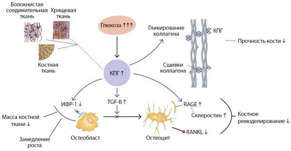
The article discusses the main mechanisms of the development of osteopenic syndrome in DM1. An analysis of domestic and foreign literature sources has shown that immune factors, impaired phosphorus-calcium metabolism as a result of chronic hyperglycemia, accumulation of glycolysis end products, decreased levels of insulin, amylin, and 25(ОН)D deficiency play a significant role in the formation of bone pathology in type 1 diabetes. In addition to the direct effect of diabetes on bone cells and the bone matrix, the risk of osteopenic syndrome is influenced by diabetic complications that occur in various body systems. Consideration of the cellular and molecular mechanisms of the formation of osteopenic syndrome associated with diabetes expands the understanding of this problem, thereby allowing not only to develop diagnostic criteria, but also to propose effective methods of prevention and treatment.
CASE REPORT
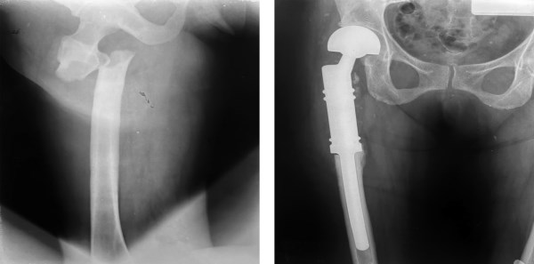
Breast cancer is the most common type of malignant disease in women. In most cases, his treatment, in addition to surgery, includes long-term use of antiestrogenic drugs that have a negative effect on bone tissue. To prevent bone loss, as well as to prevent bone metastasis, such patients are prescribed antiresorptive therapy with bisphosphonates (BP). However, there is a danger that prolonged suppression of bone remodeling processes can lead to serious complications, one of which is an atypical hip fracture. The presented clinical case describes this undesirable phenomenon in a woman with breast cancer who has been on aromatase inhibitor and BP therapy for a long time. The complex treatment, including polychemotherapy, radiation therapy, and adjuvant therapy, made it possible to achieve remission and a long relapse-free period, and the administration of zoledronic acid prevented the loss of (bone mineral density) BMD that could occur while taking letrozole. However, such prolonged use of the antiresorptive drug led to the occurrence of an atypical hip fracture followed by arthroplasty, which once again confirmed the lack of justification for long-term use of CF in a woman receiving an aromatase inhibitor. The article demonstrates the importance of careful regular monitoring of such patients, where the decision on the expediency of continuing their antiresorptive therapy should be made individually, taking into account all factors of potential risk and benefit.

This work is licensed under a Creative Commons Attribution-NonCommercial-NoDerivatives 4.0 International License (CC BY-NC-ND 4.0).
ISSN 2311-0716 (Online)


